Blood Vessels of Head and Neck
Vessels that are located on the medial aspect of the styloid process are the internal jugular vein and the internal carotid artery as well as its branches the lingual artery facial artery superficial temporal artery and the maxillary artery. Blood vessel histology Author.

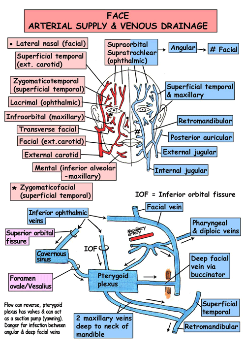
Medical Medicine Doctor Nurse Md Medic Medicalschool Medschool Paramedic Finals Exams Medschoo Medical Anatomy Anatomy Human Anatomy And Physiology
It also includes the veins that return deoxygenated blood from these organs to the heart.

. Lorenzo Crumbie MBBS BSc Reviewer. They further divide into arterioles and capillaries. This is in fact the order in which blood circulation occurs.
This technique is able to create pictures of the blood vessels in the head and neck. March 28 2022 Reading time. Dimitrios Mytilinaios MD PhD Last reviewed.
This article shall explore the anatomy of this arterial system its anatomical course branches and clinical correlations. You will be asked to lie on a narrow table that slides into the center of the CT scanner. Several important vessels lie in the vicinity of the styloid process.
A computer creates many separate images of the body area called slices. While inside the scanner the machines x-ray beam rotates around you. The head and neck receives the majority of its blood supply through the carotid and vertebral arteries.
17 minutes It would be impossible to get blood to the predestined locations without the vascular pathways. These images can be stored. Arteries of the head and neck carotids Brachiocephalic artery.
Blood vessels form the extensive networks by which blood leaves the heart to supply tissue. Arteries and veins contain three layers. The cardiovascular system of the head and neck includes the vital arteries that provide oxygenated blood to the brain and organs of the head including the mouth and eyes.
Arteries make up only a portion of the blood vessels in the body. The vessels of the body include arteries arterioles capillaries venules and veins. Blood Supply and Lymphatics.
Tunica externa outer layer - comprised of. Blood vessels of the body. How the Test is Performed.
Capillaries are the blood vessels where the exchange of oxygen nutrients and waste. Arterioles are the smallest arteries and they connect directly to capillaries to form the capillary bed. Among these blood vessels are several unique and important structures that have evolved to help.
Instant Anatomy Head And Neck Vessels Arteries Face Dental Hygiene School Medical Anatomy Dental Anatomy

Head And Neck Vessels Radiology Case Radiopaedia Org Arteries Anatomy Medical Anatomy Carotid Artery

Superficial Veins Of The Head And Neck Human Anatomy And Physiology Medical Anatomy Superficial Veins

Carotids Arteries Anatomy Medical Anatomy Anatomy Organs

This Diagram Shows The Veins Present In The Head And Neck Anatomy And Physiology Medical Anatomy Physiology

Pin On Heart Anatomy

Pin On Anatomy Physiology

Pin On Grammar

Pin On Stroke

Pin On Medical

Pin On My Work

Class Blog Bio 202 Arteries And Veins Key Arteries And Veins Medical Anatomy Arteries Anatomy
Pin On Fnp

Venous System Of Head Neck Facial Veins Cardiovascular System Ultrasound Tech

Class Blog July 2012 Arteries And Veins Arteries Anatomy Arteries

Pin On Anatomy

Carotid Artery Anatomy Chiropractic Neck Manipulation And Risk Of Stroke Carotid Artery Vertebral Artery Arteries Anatomy

Carotid Artery Anatomy Chiropractic Neck Manipulation And Risk Of Stroke Carotid Artery Vertebral Artery Arteries Anatomy

Pin On Slp


Comments
Post a Comment